![[Translate to English:]](/fileadmin/_processed_/d/8/csm_Labor_02_e8569f73ba.jpg "[Translate to English:]")
![[Translate to English:]](/fileadmin/_processed_/7/e/csm_Labor_05_5e3070a8e8.jpg "[Translate to English:]")
![[Translate to English:]](/fileadmin/_processed_/6/d/csm_Labor_07_df7204d178.jpg "[Translate to English:]")
![[Translate to English:]](/fileadmin/_processed_/1/9/csm_Labor_09_23a223fed5.jpg "[Translate to English:]")
![[Translate to English:]](/fileadmin/IAAC/images/Bilder_Labor/Labor_03.jpg "[Translate to English:]")
![[Translate to English:]](/fileadmin/_processed_/1/4/csm_Labor_04_b68a5b590d.jpg "[Translate to English:]")
![[Translate to English:]](/fileadmin/_processed_/1/5/csm_Labor_06_3df28ab249.jpg "[Translate to English:]")
![[Translate to English:]](/fileadmin/IAAC/images/Bilder_Labor/Labor_08.jpg "[Translate to English:]")
![[Translate to English:]](/fileadmin/_processed_/c/d/csm_Labor_10_87bdab6b4e.jpg "[Translate to English:]")
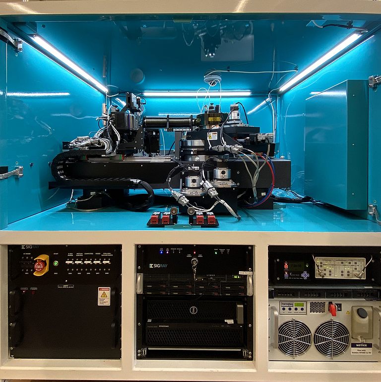
2D- & 3D-micro-XRF
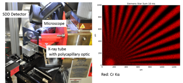
A 2D micro XRF instrument (XRF = x-ray fluorescence) has been developed in the working group at WSU. This could be further developed into a confocal 3D-micro-XRF instrument. For this purpose, polycapillary optics are used to focus the X-ray beam on a spot with a diameter of about 20 μm. An energy dispersive Silicon Drift Detector (SDD) enables the acquisition of a spectrum for each pixel (or in 3D: each voxel). Thus, information about the elemental distribution in the sample is obtained quasi-disturbance-free. This and the indifference of the method to atmospheric pressure is an advantage over electron microscopy. The comparatively greater penetration depth of the X-ray beam into the sample enables analysis at depth below the surface.
X-ray fluorescence analysis (XRF) is particularly suitable for the determination of elements with high atomic numbers. Due to the non-destructive nature of this method (excitation with photons/detection of photons), only a comparatively small amount of sample manipulation is required; for example, a battery electrode can be examined directly in an open battery. Liquid samples can be examined in a glass capillary.
CMXRF (confocal micro-XRF)
Confocal micro X-ray fluorescence analysis provides an exclusive view below the surface. Using synchrotron radiation at the ANKA-FLUO-beamline at KIT (Karlsruhe, Germany), we have obtained 3D elemental distributions in anodes of aged lithium ion batteries. These measurements will soon also be possible in our laboratory.